
Abstract
Objective
Pineal region tumors are frequently large at diagnosis, often presenting with obstructive hydrocephalus. Surgical resection can be challenging due to intrinsic tumor vascularity and the surrounding eloquent neurovascular anatomy. For patients with large pineal region tumors, particularly young children, neoadjuvant chemotherapy may decrease both the size and vascularity of the tumor before definitive resection. Herein we present our experience.
Methods
A single-center retrospective review of pediatric patients with pineal region/posterior third ventricular tumors was performed. Each patient received at least 2 rounds of neoadjuvant chemotherapy (carboplatin, etoposide, cyclophosphamide) prior to definitive resection. The primary outcome measure for this study was the radiographic effect of said chemotherapy on the tumor. Pre- and post-chemotherapy tumor volumes were calculated using magnetic resonance imaging tumor volume analytics. Secondary outcomes were estimated intraoperative blood loss, extent of resection, operative complications, readmission rates, and patient survival. Blood loss was calculated as a percentage of estimated total blood volume.
Results
We identified thirty-one children and young adults (16 males, 15 females) who underwent neoadjuvant chemotherapy for pineal region tumors. The mean age of the population was 7.42 years (5 mo–22 yr). Pathologies were predominantly embryonal and included pineoblastoma (n=22; 71%), atypical teratoid rhabdoid tumor (n=5; 16%), pineal parenchymal tumor of intermediate differentiation (n=3; 10%), and embryonal tumor with multilayered rosettes (n=1; 3%). The average percent tumor volume reduction post chemotherapy was 51% (range, -12.4–90.0%); only 1 patient had an increase in tumor volume. There were no complications related to chemotherapy. All surgeries resulted in gross total or near total resections. Average blood loss as a percentage of preoperative total blood volume was 11.7% (1.1–38.8%). Average length of follow-up was 6.33 years (range, 1–14 years) with 20 patients (64.5%) alive at last follow-up.
Conclusions
Neoadjuvant chemotherapy for certain pineal region tumors is a safe and effective means to decrease tumor volume. While we believe this reduction in tumor size facilitates subsequent resection, further multicenter studies are needed.
Introduction
Tumors of the pineal region are relatively rare, representing 3–11% of all pediatric brain tumors [1,2,3]. Germ cell and pineal parenchymal tumors are the main lineages found in children [2]. Patients commonly present with obstructive hydrocephalus but may also have symptoms secondary to mass effect on surrounding structures, such as the dorsal midbrain or cerebellum. These tumors, particularly in young children, may be quite large and vascularized at the time of diagnosis [2, 4].
Pineal tumors in children require a multimodal approach integrating clinical, radiographic, and laboratory data [5]. High resolution magnetic resonance imaging (MRI) defines the tumor size, extent, and relationship to surrounding venous anatomy, while cerebrospinal fluid (CSF) and serum tumor markers (alpha-fetoprotein [AFP], beta human chorionic gonadotropin [β-hCG]) may guide the diagnosis and obviate the need for a surgical biopsy in select cases [2]. If hydrocephalus is present, intraventricular endoscopy may provide a minimally invasive means to treat the hydrocephalus via endoscopic third ventriculostomy (ETV), which provides an opportunity to concurrently obtain CSF +/- tumor tissue for analysis [6, 7]. However, there are known and suspected risk factors for ETV failure such as young age, presence of metastatic disease, and malignant pathology [8,9,10]. Patel et al. reported a single center ETV failure rate of 42.3% in pineal tumor patients [11]. Alternatively, if hydrocephalus is not present, a lumbar puncture and craniotomy may be needed to obtain CSF and tumor tissue, respectively.
Management of pineal region tumors remains complex because of their histologic heterogeneity and variable response to chemotherapy and radiotherapy, notably in young children where treatment-related toxicities pose significant challenges [12, 13]. Gross or near total resection has been correlated with improved survival in certain tumor types without metastatic disease, as well as in a subset of children with metastatic disease [4, 14]. However, the deep-seated location and surrounding neurovascular structures of the pineal region make it demanding to access surgically. This can be compounded by large tumor size and hypervascularity [15]. To address these challenges, multidisciplinary, patient-tailored treatment strategies have been emphasized [13, 14].
Pre-resection (i.e., neoadjuvant) chemotherapy can reduce tumor vascularity, induce necrosis and cystic changes, and reduce overall tumor volume, thereby potentially improving the safety and feasibility of subsequent surgical resection. This strategy has been used effectively in choroid plexus carcinoma (CPC) and embryonal tumors [4, 16]. However, its utility for pineal region tumors has not been well characterized. Herein, we present our experience with neoadjuvant chemotherapy prior to definitive resection of pineal region tumors in a predominantly pediatric population.
Methods
Study design and population
Institutional review board approval was granted by the University of Tennessee Health Science Center, Memphis; patient consent was waived because of the retrospective design of the study. Patients were identified from the brain tumor database at Le Bonheur Children’s Hospital, Memphis, TN from 2015 to 2025. All patients who underwent resection of pineal region tumors were screened. We searched the electronic medical record system to determine age, tumor pathology, and whether patients underwent neoadjuvant therapy. Inclusion criteria were children and young adults with a confirmed tumor of the pineal region who received at least 2 rounds of neoadjuvant chemotherapy prior to attempted resection. Patients with prior partial resection or biopsy performed at an outside facility were included. As a group, germ cell tumors were excluded from this study as resection is not a standard part of their treatment (e.g., germinomatous and non-germinomatous germ cell tumors) or chemotherapy is ineffective (e.g., teratoma). We recognize that some pineal germ cell patients will undergo so-called 2nd look surgery to remove post-treatment residual abnormalities, but this is rare.
Data collection
Demographic information, follow-up details, and pre-, intra-, and post-operative data were collected. The primary outcome of this study was the change in tumor volume after neoadjuvant chemotherapy. Additional outcomes were intraoperative blood loss, extent of resection (EOR), operative complications, readmission rates, and patient survival. Gross total resection (GTR) was defined as no demonstrable residual tumor on MRI; near total resection (NTR) was defined as > 90% tumor resection and subtotal resection (STR) was < 90%. Blood loss was calculated as a percentage of estimated total blood volume.
Tumor volumetric analysis
Pre- and post-chemotherapy contrast-enhanced brain MRIs were uploaded onto a segmentation platform (Mint Medical, Hamilton NJ). Pineal tumors were segmented on axial 2D or 3D post-contrast T1sequences using a 3D segmentation tool by a board-certified radiologist with expertise in pediatric neuroimaging (SP). Tumor volumes, bi-perpendicular diameters and their percentage changes were automatically generated by the software.
Treatment algorithm
Treatment begins with CSF diversion, if needed, and analysis of serum and CSF germ cell markers. When hydrocephalus is present, an ETV and concurrent endoscopic tumor biopsy is preferred, but a risk-benefit analysis needs to be undertaken first if the surgeon has concerns regarding the risk of tumor bleeding. If endoscopic biopsy was not performed or was non-diagnostic, then an open biopsy via craniotomy is strongly considered. Tissue diagnosis is always preferred prior to starting neoadjuvant chemotherapy, but in some circumstances, we will proceed without pathologic diagnosis (e.g., bleeding complication with prior non-diagnostic biopsy or imaging features that make the surgeon feel that biopsy-induced bleeding is a high risk).
All patients are discussed at a multidisciplinary tumor board. Neoadjuvant chemotherapy is considered in all patients, but particularly those with the following features: (1) imaging and pathology that show significant tumor vascularity; (2) large tumor size; and (3) young age that increases the risk of excessive blood volume loss. There are no specific age or tumor size parameters; these and other factors are collectively used to formulate a treatment recommendation. The goal of neoadjuvant chemotherapy is to reduce the size and vascularity of the tumor, and alter the consistency (e.g., necrosis) and composition (e.g., cystic transformation). Patients typically undergo 2 rounds of chemotherapy. Each round of chemotherapy typically proceeds as follows: day 1 includes carboplatin 560 mg/m2 IV and etoposide 100 mg/m2 IV; day 2 includes cyclophosphamide 1200 mg/m2 IV with mesna support and etoposide 100 mg/m2 IV [17, 18]. On day 3, granulocyte colony stimulating factor (5mcg/kg) is given for 10–14 days. Each cycle is 3–4 weeks in total duration. After 2 cycles of chemotherapy, the patient’s case is re-presented at our multidisciplinary conference where pre- and post-chemotherapy imaging is reviewed. A decision is made whether to continue with additional chemotherapy or proceed with definitive resection. Time from completing chemotherapy to surgery averages about 2 weeks, being primarily a function of recovery of blood counts and operating room availability.
Results
Demographic information and prior surgeries
A total of 31 patients with pineal region tumors underwent neoadjuvant chemotherapy followed by tumor resection (Table 1). There were 16 (51.6%) males and 15 (48.4%) females with a mean age on the date of surgery of 7.42 years (range, 5 months–22 years). Twenty-nine patients were under the age of 18, but there were 2 patients older than 18 years (19 and 22-year-old). Most patients were Caucasian (71.0%). Eleven (35.5%) patients had an existing shunt, 21 (67.7%) patients had a prior ETV, 25 (80.6%) patients had a prior biopsy, and 3 (9.7%) patients had a prior subtotal resection. Three patients did not have pathologic diagnosis prior to resection as it was felt there was a significant risk of bleeding with endoscopic biopsy; the imaging in these 3 patients were suggestive of an embryonal-type tumor.
Tumor pathology and volumetric analysis
The most common type of tumor was pineoblastoma (n = 22; 71.0%), followed by atypical teratoid/rhabdoid tumors (ATRT) (n = 5; 16.1%), pineal parenchymal tumors of intermediate differentiation (PPTID) (n = 3; 9.7%), and embryonal tumors with multilayered rosettes (ETMR) (n = 1; 3.2%) (Table 2). Most patients received 2 rounds of neoadjuvant chemotherapy (n = 27, 87.1%); the remaining (n = 4, 12.9%) received 3 rounds. There were no documented complications related specifically to neoadjuvant chemotherapy, including infectious or hemorrhagic complications.
Mean pre-chemotherapy tumor volume was 24.7 cm3 (range, 1.1–84.1 cm3), which decreased on average by 51.0% to 12.1 cm3 (range, 0.5–54.5 cm3) following chemotherapy (Table 2). This decrease resulted in an average absolute tumor volume reduction of 12.6 cm3 (range, -6.0–65.5 cm3). ATRTs (n = 5) were the largest tumors by volume on the initial MRI with a mean volume of 39.5 cm3 (range, 1.1–84.1 cm3), followed by ETMR (n = 1) at 30.1 cm3 (no range), pineoblastomas (n = 22) at 22.1 cm3 (range, 3.5–81.5 cm3), and PPTID (n = 3) at 16.6 cm3 (range, 5.0–37.3 cm3). ETMR, ATRTs, and pineoblastomas were the most responsive to chemotherapy with a 56.9% (no range), 54.7% (range, 10.53–74.5%), and 49.8% (range, -12.4–84.2%) volume reduction, respectively. PPTIDs had less reduction in tumor volume on average at 41.2% (range, 28.4–90.0%). Figure 1 shows the change in tumor volume for all 31 patients by tumor type.
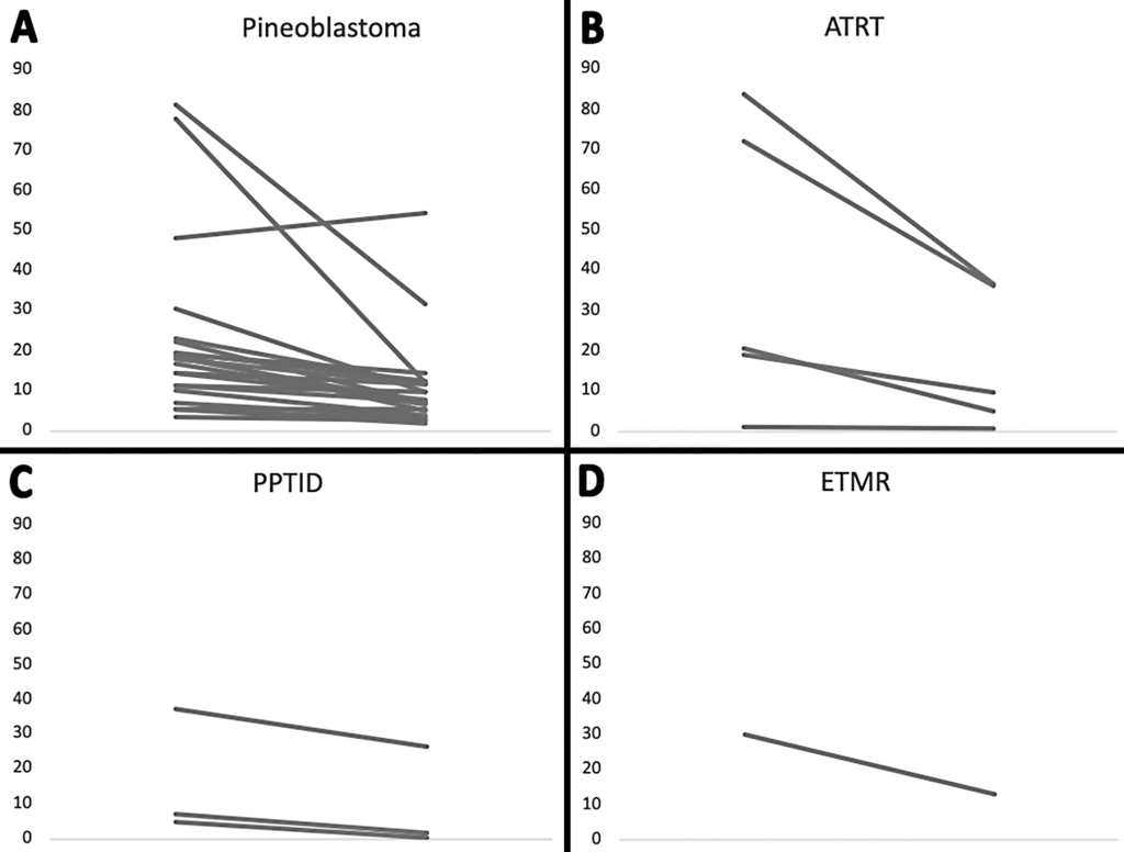
Slope plots depicting the change in tumor volume pre- and post-chemotherapy of all 31 patients. Panels A, B, C, and D show the tumor volume change in pineoblastomas, ATRTs, PPTIDs, and ETMRs, respectively. Patient 5 in Panel A was the only patient with tumor growth during the neoadjuvant chemotherapy course
Only 1 patient showed tumor growth on chemotherapy—a 16-year-old boy with a large pineoblastoma/pineal anlage tumor (PAT) who exhibited a 12% increase in tumor volume after 3 cycles of chemotherapy. Maximum absolute tumor reduction after chemotherapy was 65.6 cm3 (pre-chemotherapy volume of 77.9 cm3 and post-chemotherapy volume of 12.3 cm3) in a 22-year-old patient with pineoblastoma (Fig. 2).
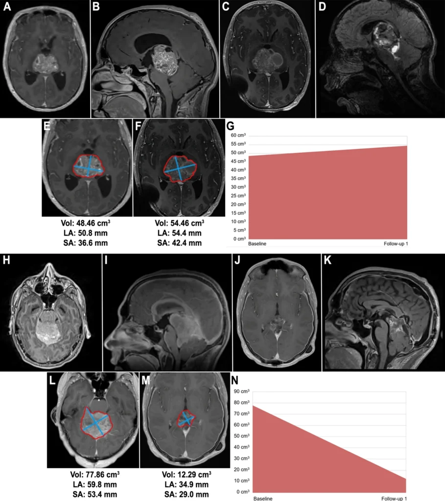
Case 1. Imaging of the only patient in our series whose tumor progressed on chemotherapy. This was a 16-year-old male with a PAG. Pre-chemotherapy (A) axial, and (B) sagittal T1-weighted with contrast MRI demonstrating the pineal mass. Post-chemotherapy (C) axial, and (D) sagittal T1-weighted with contrast MRI imaging demonstrating an increase in the volume of the mass. MRI tumor analytics in (E) and (F) show the calculated volume (cm3), long axis (mm), and short axis (mm) of the mass. (G) Visual depiction of an increase in tumor volume by 12%. Case 2. Imaging of a 22-year-old male with a pineoblastoma that decreased in size during pre-operative chemotherapy. Pre-chemotherapy (H) axial, and (I) sagittal T1-weighted with contrast MRI demonstrating a pineal mass. Post-chemotherapy (J) axial, and (K) sagittal T1-weighted with contrast MRI imaging demonstrating a decrease in the volume of the mass. MRI tumor analytics in (L) and (M) showing the calculated volume (cm3), long axis (mm), and short axis (mm) of the mass. (N) Visual depiction of a decrease in tumor volume by 65 cm3 (84%), the maximum absolute tumor volume reduction in our series
Surgical data: operative time and extent of resection
The mean operative time was 6.2 h (range, 2.9–10.5 h). A total of 20 (64.5%) GTRs were achieved: all ATRTs (n = 5; 100.0%), the lone ETMR (n = 1; 100%), 12 of the 22 pineoblastomas (54.5%), and 2 of the 3 PPTIDs (67%). The remaining 11 cases (35.5%) were NTRs (Table 1).