
Abstract
Surgical resection for medulloblastoma has been a mainstay of treatment for decades due to the need for definitive diagnosis and known benefits on treatment efficacy and survival. While many pediatric neurosurgeons and neurooncologists agree that maximal safe surgical resection should be the goal of medulloblastoma surgery, actual practical application and benefits of maximal safe resection seem to vary. Early work on extent of resection provided a relatively arbitrary 1.5 cm2 threshold to distinguish subtotal resection (STR) from near total resection (NTR) and demonstrated a survival benefit if the residual was ≤ 1.5 cm2 (NTR or gross total resection (GTR)). However, recent evidence questions the validity of this threshold, with European researchers even suggesting that STR alone should not be considered a high-risk feature during adjuvant therapy planning. The question regarding extent of resection, therefore, is undeniably controversial and has become even more complex in recent years due to the seminal work done to identify medulloblastoma subgroups based on molecular characteristics. Adjuvant treatment approaches are being tailored to these molecular subgroups, and the impact of extent of resection on survival is being reconsidered within the context of subgroups. Certainly, the goal when treating children with medulloblastoma, or any form of brain cancer, should be to cure the disease while minimizing treatment-related toxicity, including surgical injury. In this article, we discuss the role of surgical resection for treating medulloblastoma and whether it is possible to maximize both surgical resection, to potentially improve survival and adjuvant treatment efficacy, while minimizing neurological morbidity.
Keywords: Extent of resection · Medulloblastoma · Near total resection
1 Surgical Neurology Branch, NINDS, National Institutes of Health, Bethesda, MD, USA
2 College of Medicine, University of Tennessee Health ScienceCenter, Memphis, TN, USA
3 St. Jude Children’s Research Hospital, Memphis, TN, USA
4 Division of Neurosurgery, Connecticut Children’s, Hartford,CT, USA
5 Department of Neurosurgery, UConn School of Medicine,Farmington, CT, USA
6 Department of Neurosurgery, University of Tennessee HealthScience Center, Memphis, TN, USA
7 Neuroscience Institute, Le Bonheur Children’s Hospital,Memphis, TN, USA
8 Semmes Murphey Clinic, Memphis, TN 38120, USA
9 Department of Neurosurgery, Great Ormond Street Hospitalfor Children, London, UK
History of surgical resection for medulloblastoma
Medulloblastoma is the most common malignant primary brain tumor in children [18]. It is estimated that approximately one-third of children with medulloblastoma eventually succumb to their disease, while those who survive often experience significant morbidity related to disease progression and treatment effects [23]. The Children’s Cancer Group 921 study (CCG-921) was conducted in the 1980 s and 1990 s to examine the correlation between extent of medulloblastoma resection and prognosis in 188 patients. In addition to overall survival (OS) benefit for older children (older than 3 years) and patients with non-metastatic disease, CCG-921 demonstrated a progression-free survival (PFS) advantage for lower residual tumor burden [1, 31]. The study established 1.5 cm2 as the threshold for residual tumor that distinguishes near-total resection (NTR) from sub-total resection (STR), based predominately on CT imaging. For the first time, a quantifiable imaging-based measurement was put forth as a “gold standard” for the evaluation of surgical resection, rather than relying on the often inaccurate intraoperative observations of the neurosurgeon. However, this threshold was not established by an a priori hypothesis but instead was reached through an iterative statistical process that resulted in statistically significant separation of the survival curves [1, 31]. The authors noted that residual tumor ≤ 1.5 cm2 on postoperative scans could significantly improve PFS, likely because a smaller tumor burden is more amenable to effective control by adjuvant therapy. Other subsequent studies—such as the International Society of Pediatric Oncology (Société internationale d’oncologie pédiatrique or SIOP) Primitive Neuroectodermal Tumors study (PNET 4, 2012) which compared hyperfractionated radiotherapy with standard radiotherapy and the Children’s Oncology Group (COG) Phase III trial (ACNS033, 2021) which evaluated reduced dose and volume radiotherapy with chemotherapy—corroborated that residual disease in excess of 1.5 cm2 was associated with a worse outcome [13, 15].
Since the above-mentioned trials were conducted, this controversial threshold has remained the standard for clinical decision-making. However, the limitations of these trials coupled with potentially greater morbidity from more aggressive resections have led some groups to question whether the 1.5 cm2 “rule” retains its value in the new era of molecular subgroups and targeted therapies [28].
Medulloblastoma molecular subgroups
Since the conclusion of the CCG-921 trial in 1992, there have been many major advances in the field of neurosurgical oncology, particularly for medulloblastoma. Rapid advances in magnetic resonance imaging (MRI) technology coupled with the subspecialization of pediatric neuroradiologists have dramatically improved the imaging evaluation of medulloblastomas. Surgical techniques, instruments, and pediatric neuroanesthesia continue to be refined. Intraoperative technology, such as ultrasound and intraoperative MRI, has provided surgeons with the means to assess extent of resection in real time. Importantly, the medulloblastoma community experienced a seismic shift in 2012 with the landmark publication by Taylor et al. that delineated medulloblastomas into subgroups based on transcriptional profiling, genetic mutation patterns, and clinical characteristics rather than relying solely on histologic features [26]. The initial medulloblastoma subgroups included wingless/INT1-activated (WNT), sonic hedgehog-activated (SHH), group 3, and group 4. The most recent iteration of the World Health Organization (WHO) Classification of Tumors of the Central Nervous System (CNS) in 2021 utilized distinct clinical and biological differences, and incorporated wide-scale methylation profiling results, to develop the current state of medulloblastoma classification: medulloblastoma, WNT-activated; medulloblastoma, SHH-activated TP53 wildtype; medulloblastoma, SHH-activated TP53 mutant (further divided into 4 SHH subgroups based primarily on methylation); and medulloblastoma, non-WNT/non-SHH (formerly groups 3 and 4, now further divided into 8 subgroups) [14, 24].
While further changes are likely to occur in future classification systems, medulloblastoma subgroups have now become integrated into prognostication, as well as in the development and application of targeted, risk-stratified therapeutic strategies [3, 5, 16, 25]. Despite the explosion of molecular information, the specific impact of extent of surgical resection on each individual subgroup is largely unknown. An unintended consequence of the molecular classification of medulloblastoma—already a relatively rare tumor—into multiple subgroups and subtypes is that the number of patients within each individual classification becomes smaller and smaller, which makes determining the effect of any intervention, including surgery, harder to evaluate. Furthermore, potential confounders, such as age and the presence of metastatic disease, may also contribute disproportionately from group to group. As a result, specific clinicopathological characteristics and the natural history of individual subgroups may affect the risk-benefit analysis for surgery. For example, given their excellent prognosis, children with medulloblastomas in the WNT subgroup may not require the surgical resection to be as aggressive as for medulloblastomas in other subgroups. However, in patients with a newly diagnosed posterior fossa tumor, preoperative imaging may not even accurately predict the general histopathological diagnosis of medulloblastoma, let alone the specific molecular subgroup. Therefore, information regarding the medulloblastoma subgroup is currently unable to influence surgical decision-making for a primary resection. This issue may be settled in the future with ongoing advances in nanopore sequencing, which facilitates a rapid intraoperative diagnosis [4, 8, 19]. Ultimately, such technology coupled with effective non-operative therapy for specific molecular subgroups may allow a paradigm shift to biopsy or STR (coupled with hydrocephalus management). Until then, however, surgical planning must proceed in the absence of molecular data.
Ongoing controversy—different strategies based on institution and region
While our molecular knowledge of medulloblastoma has advanced tremendously, the question of the role of resection has become ever more complicated and vexing [21]. As we move closer to having longer survival data available for patients treated in the molecular subgroup era with different targeted strategies, it appears we are currently at a crossroads concerning the extent of surgical resection in pediatric medulloblastoma patients—do surgeons strive for maximal resection, ideally with a residual of less than 1.5cm2, or do they debulk or even just biopsy and proceed with adjuvant therapy? This dilemma is highlighted not just by alternative surgical opinions from the literature across different geographic locations, but also by a poll conducted at the 2020 joint American Association of Neurological Surgeons/Congress of Neurological Surgeons (AANS/CNS) Pediatric Section Meeting. The audience was asked what their preferred intervention would be for a pediatric patient found to have a 2.0 cm residual lesion in the right cerebellomedullary angle (Fig. 1A). An almost equivalent number of attendees responded that they would reoperate versus leaving the residual and starting adjuvant therapy (Fig. 1B).
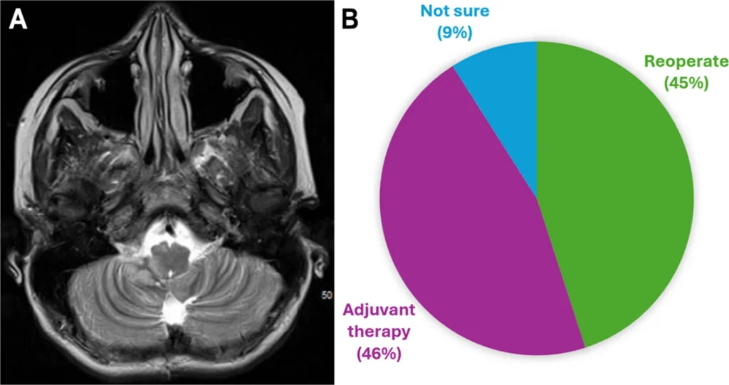 Fig.1
Fig.1
A A small residual medulloblastoma (2.0 × 1.4 cm) in right cerebellomedullary angle. B Summary of responses to a poll administered at the 2020 AANS/CNS Pediatric Section Meeting that asked respondents about their preferred treatment strategy for such a lesion
A retrospective study by Thompson et al. of 787 patients with medulloblastoma treated at 35 international institutions found a PFS benefit when comparing gross total resection (GTR) with STR but found no differences in PFS or OS when comparing GTR with NTR [28]. Furthermore, when analyzing based on subgroups, only the group 4 subgroup had a PFS survival advantage after GTR. The authors argued that extent of resection for certain subgroups could be limited to avoid morbidity and suggested leaving residual disease behind that is adherent to critical structures. It should be noted that extent of resection was based on non-centralized, institution-specific radiologist reads of postoperative T1 MRIs. In addition to the retrospective study, Thompson et al. undertook a systematic review of studies related to extent of resection in medulloblastoma and found similar studies supporting (n = 16) or refuting (n = 20) an association between extent of resection and survival [27]. Fourteen studies had mixed results, while only three articles included molecular subgroup analysis. There were significant limitations in the 50 studies that were included in their analysis. Articles were published across a 40-year time span, from 1977 to 2017. All articles were retrospective either in analysis alone or in both data collection and analysis. There was no consensus on how extent of resection was assessed or categorized, including some that were based solely on surgeons’ reports. Given the results of these two important studies by Thompson and colleagues, what is clear is that there is no definitive answer about the prognostic significance of extent of resection.
In the USA and Canada, children with medulloblastoma are often considered high-risk if they present with metastases at diagnosis, are younger than 3 years of age, and/or have residual disease ≥ 1.5 cm2. We surmised from the findings of the CCG-921 study, as well as from collective experience with other brain tumors in which increased extent of resection generally yields improved survival, that achieving a GTR/NTR when safely feasible is still the surgical goal in medulloblastoma at most high-volume pediatric institutions in North America. Depending on the institution, some high-risk medulloblastoma patients with unequivocal or suspected residual tumor (especially if the residual tumor is ≥ 1.5 cm2) may undergo a second-look surgery or be treated according to high-risk protocols with intensified chemoradiotherapy regimens [20, 28].
In recent years, researchers in the UK and Europe have questioned the validity of STR (i.e., > 1.5 cm2 of residual tumor) as a high-risk disease feature. In fact, Simon Bailey and coauthors state that, “extent of surgical resection is no longer considered a prognostic variable in medulloblastoma,” citing several primarily European studies [2]. For example, in the European SIOP High-Risk Medulloblastoma (SIOP-HR-MB) clinical trial for non-infant, high-risk medulloblastoma, STR was not considered a high-risk feature in the absence of other high-risk features: metastatic disease, large cell/anaplastic histology (LCA), MYC amplification, TP53 amplification, or MYCN amplification in SHH tumors [2, 11]. In their retrospective cohort of 1100 patients (416 from UK Children’s Cancer Center, and the remainder from previously published cohorts), Keeling et al. found that STR (n = 226, 20%) predicted lower overall survival compared with GTR (n = 884, 80%) [11]. However, in their multivariable analysis, STR was not found to be an independent prognostic factor across all subgroups when examined alongside other high-risk features. This led to the assertion that patients with STR should be considered as standard risk (and not high risk) for adjuvant treatment planning and clinical trial inclusion purposes [11, 28]. While this UK analysis was said to be independent of adjuvant treatment effects, it is highly likely, as noted by the authors, that the majority of STR patients underwent more intensive treatment regimens (since STR was considered a high-risk feature at the time), thus potentially complicating the findings. Furthermore, STRs in the UK cohort were more likely to be encountered in younger patients and non-WNT patients. This higher preponderance of STR in patients who were already at higher risk of poor outcomes could serve as a confounder. Lastly, a previous study by the same group demonstrated improved overall survival among patients who underwent re-resection at the time of relapse [10]. The demonstration of a survival advantage for re-resection at relapse might be extrapolated to the primary resection as well, suggesting a benefit of more aggressive resections rather than STR. We await the results from the SIOP-PNET5-MB trial, a European trial (concluded in 2022) that used clinical, histological, and molecular parameters for children and adolescents with standard-risk medulloblastoma [17].
Another surgical opinion stems from Egypt. Enayet et al. retrospectively studied 405 patients with medulloblastoma treated in Egypt [7]. The 5-year overall survival was reported to be 79.5% for STR versus 87.6–96.3% for GTR/NTR, while the 5-year PFS was 70.1% for STR versus 83.9–86.1% for GTR/NTR. Ultimately, the authors found no significant OS (p = 0.557) or PFS (p = 0.146) benefit between GTR/NTR or STR. The study also found no association between GTR and increased risk of complications, like posterior fossa syndrome; the authors therefore concluded that GTR should still be pursued whenever possible. Of note, these patients were not stratified by molecular subgroup, but age and disease dissemination were considered. The group at Dana Children’s Hospital in Tel Aviv has recently offered some thought-provoking results in patients presenting with metastatic disease [6]. Twelve children with a mean age of 6.5 years (range, 1.1–16.1) underwent biopsy only (with or without shunt), followed by adjuvant therapy. Three children under the age of 3 were treated with chemotherapy only; the remaining 9 patients had craniospinal radiation with chemotherapy. At last follow-up (median, 3.2 years), 9 (75%) were alive with the following molecular subgroups: SHH (n = 4), group 3 (n = 2), group 4 (n = 2), and unknown (n = 1). Estimated 5-year survival was 65%. Of the 3 children who died, 2 had MYC-amplified group 3 tumors and 1 was unknown. The authors concluded that this minimalistic approach should be considered in these high-risk patients.
The St. Jude experience
At the present, the philosophy at St. Jude Children’s Research Hospital (SJCRH) has been to strive for GTRs for all medulloblastoma patients, when possible, even those with mild-to-moderate metastatic disease. Our surgical goal is to achieve no measurable disease at the primary site prior to adjuvant therapy. Since many medulloblastoma patients come to SJCRH after their initial resection, about a third or more undergo second-look surgery for residual tumor. There are several scenarios where we would likely not take a patient back to the operating room for residual tumor: (1) heavy/bulky multifocal metastatic disease; (2) small amount of highly favorable (e.g., WNT) or highly unfavorable (e.g., SHH TP53 mutant, group 3 MYC amplified) molecular subgroups; (3) high risk of causing a significant and potentially irreversible neurologic deficit (e.g., tumor infiltration into eloquent structures like the facial colliculus, dentate nucleus or cerebellar peduncles); and (4) if the patient’s neurologic function was severely compromised from the initial resection (e.g., severe posterior fossa syndrome). Additionally, in some cases, it may be reasonable to perform a planned staged approach whereby an immediate decompression of the brainstem and opening of the CSF pathways is performed first, followed later by a second-look surgery. Second-look surgery may sometimes occur after neoadjuvant chemotherapy to shrink the residual tumor. It should be emphasized that each patient is thoroughly vetted in a multidisciplinary fashion.
Postoperative changes, blood products, and hemostatic agents can make determination of residual disease quite difficult. In a study of our experience with performing second-look surgery for medulloblastoma, we found that the overwhelming majority of patients had histopathologically confirmed residual tumor identified during the second-look surgery, even when the initial postoperative scan was indeterminate [20]. The most common sites of residual tumor were in lateral regions (lateral recess and/or foramen of Luschka, 67%) and the fourth ventricular roof (superior medullary velum, 76%).
The St. Jude Treatment of Patients With Newly Diagnosed Medulloblastoma, Supratentorial Primitive Neuroectodermal Tumor, or Atypical Teratoid Rhabdoid Tumor (SJMB03) prospective trial, performed from 2003 to 2011 and involving 330 patients with medulloblastoma, found that extent of resection remained a significant clinical risk factor. Out of the 330 patients, 315 (95.5%) had either a GTR (n = 246) or NTR (n = 69); only 13 patients had STR and 1 patient had biopsy. In comparison, the retrospective UK study by Keeling et al. (n = 1110) had a GTR rate of 80% (884/1110) with the remaining patients (n = 226) had STR. The St. Jude study demonstrated that average-risk patients (including those with NTR) achieved 5-year PFS of 83.2%, while high-risk patients (including those with prior STR) had a 5-year PFS of 58.7% (Fig. 2A) [9]. For the overall cohort in the St. Jude study, the 5-year PFS and overall survival were 75.6% and 82.3%, respectively. In the UK study, the 5-year overall survival was 67.6%. Because extent of resection was a major driver of risk classification, the SJMB03 trial highlights that maximal safe resection may alter a patient’s overall risk due to lower disease burden and less potential therapeutic side effects from less intensive adjuvant therapy regimens, while also leading to more favorable outcomes (Fig. 2B). Similarly, the St. Jude Risk-Adapted Therapy for Young Children with Medulloblastoma (SJYC07) study, a Phase II trial for children less than 3 years old from 2007 to 2017, incorporated extent of resection, among other features, into the risk-stratification criteria [22]. The low-risk group contained patients with a GTR or NTR (defined as a residual of < 1cm2) whereas the intermediate group had STR. Of the 81 enrolled patients, 68 (84%) had GTR or NTR; the rest (16%) had STR. While event-free survival did not improve compared to historical controls in this trial, the low risk group did display improved survival over intermediate and high-risk groups.
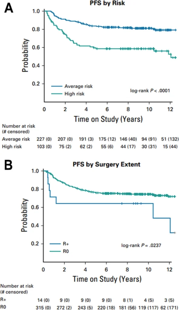 Fig.2
Fig.2
Progression-free survival based onArisk andBextent ofresection (R+=STR; R0=GTR/NTR). Reproduced with permissionfrom Gajjar A. et al. (J Clin Oncol, 2021) [9]
Maximizing resection and minimizing risk—surgical strategies
Optimizing surgical technique, utilizing available intraoperative technology (e.g., intraoperative MRI, intraoperative ultrasonography), and incorporating close neuromonitoring can help mitigate the risk of neurological morbidity even during more aggressive resections. A feared but unfortunately still common neurologic morbidity associated with GTR or NTR of posterior fossa tumors is cerebellar mutism syndrome (CMS)/posterior fossa syndrome (PFS). This syndrome is characterized by reduced or absent speech, emotional and behavioral lability, cerebellar dysfunction, dysphagia, and other motor signs, typically developing between 48 and 72 hours postoperatively. It is a direct result of manipulation of the proximal efferent cerebellar pathway (pECP) [29]. In some studies, CMS is estimated to affect at least 20% to 40% of children following medulloblastoma resection [30]. While there is evidence of certain non-modifiable risk factors, such as young age, a large midline tumor, and medulloblastoma pathology, there are surgical strategies and techniques that can reduce the risk of CMS.